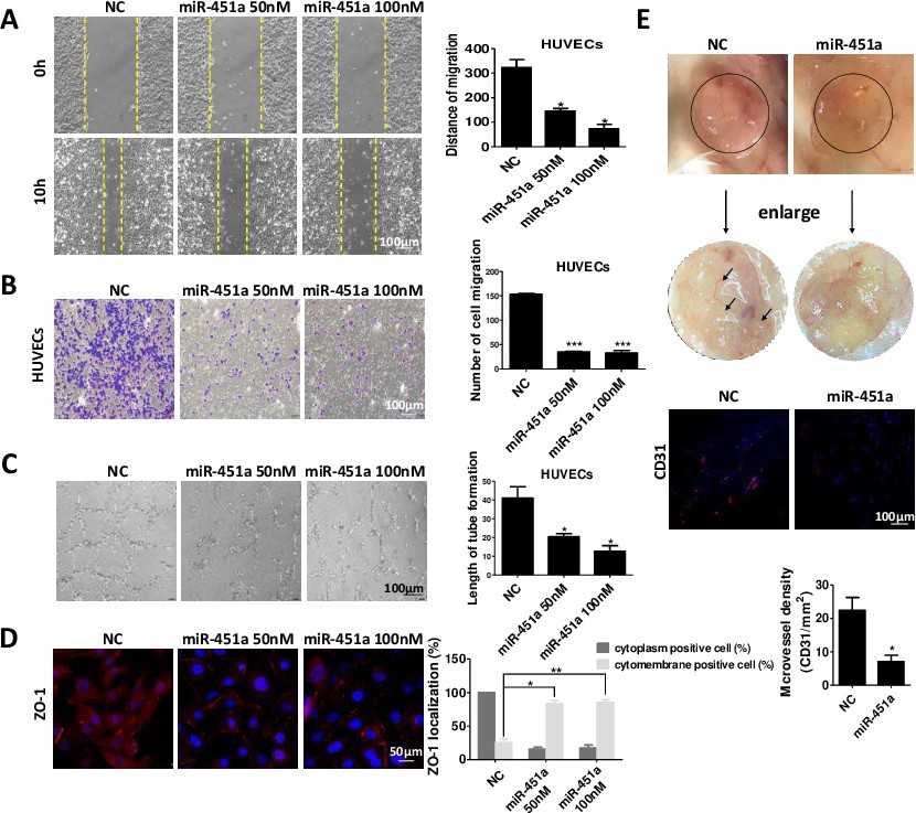
Fig. 4. miR-451a inhibits endothelial cell function. A-B, Different concentrations of miR-451a mimics were transfected into HUVECs. Cell migration was determined by wound-healing (A) and transwell (B) assays. C, HUVECs transfected with different concentrations of miR-451a mimics were seeded on a Matrigel layer. After 4 h, tubular structures were manually counted. D, Representative images showing ZO-1 staining in HUVECs transfected with different concentrations of miR-451a mimics. ZO-1-positive staining in the cytoplasm or cytomembrane was quantified, and results are presented as the percentage of positive cells. E, Matrigel containing 5 nmol miR-451a agomir or control (NC) was subcutaneously injected into mice, and plugs were surgically removed after 10 days. The expression of CD31 in plugs was examined by immunofluorescent staining. NC, negative control; *P<0.05, **P<0.01, ***P<0.001.